



01 / X-RAY
Weight-bearing X-ray
The starting point for joint space and leg alignment.
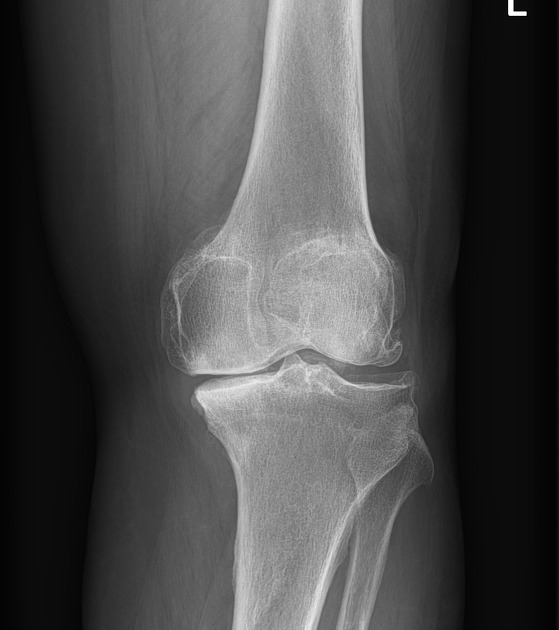
Standing X-ray of the knee. Example image for illustration; marker positions are approximate.
घुटना बदलवाने से पहले, उसे बचाने की सोचिए।
A single Subchond injection may delay the need for knee replacement. In two long-term studies, 80–82% of treated knees had not been replaced at about 15 years.
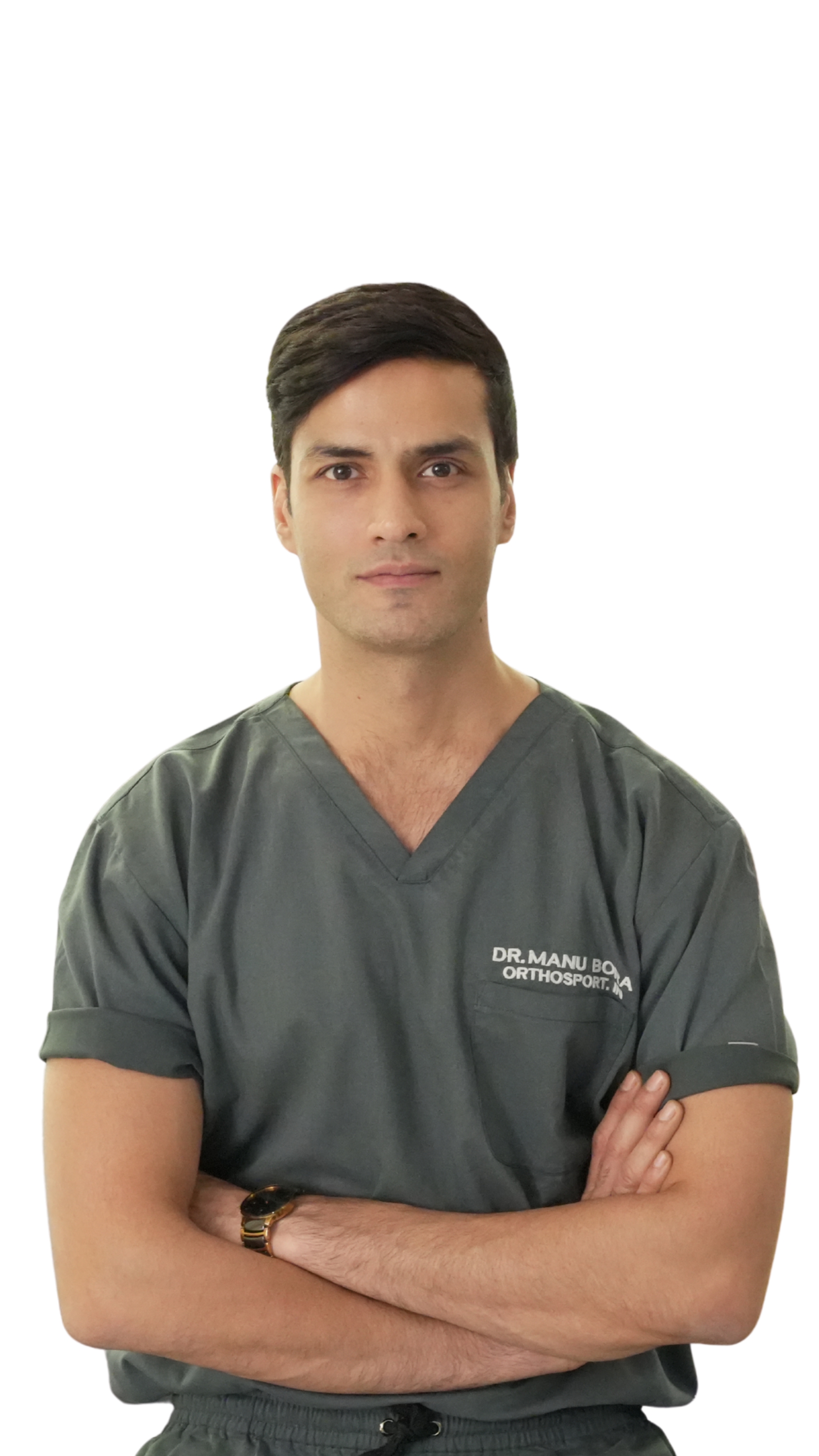
Arthrex Master Instructor · Sports Medicine & Arthroscopy
80–82%
of treated knees had not been replaced at about 15 years of follow-up.
2
independent long-term studies reported the same joint-preservation outcome.
1
Subchond injection, performed as a joint-preservation option.
Outcomes are drawn from published follow-up studies and may not reflect individual results. A clinical assessment is needed to determine suitability.
Sports Medicine & Arthroscopy Specialist
Subchondral & Natural Knee Longevity
Gurugram · New Delhi · Mumbai
Personalized Non-Surgical & Surgical Plans
Osteoarthritis is a whole-joint condition. Cartilage has little to no direct pain sensation; other tissues can contribute to pain, stiffness and loss of function.
Not only: “How much cartilage is left?” But also: “What is driving my pain?”
Hover a factor or use the arrows to see where it acts in the joint.
01Subchondral bone stress
The living, vascular, pain-sensitive bone directly beneath cartilage can undergo overload and remodeling.
Illustrative anatomy — where each factor acts
Dr. Manu Bora is an orthopaedic surgeon specialising in treating sports injuries and early joint damage with arthroscopic (keyhole) surgery and biological procedures designed to protect the bone and cartilage you already have.
He teaches arthroscopic techniques to other surgeons, and trained in France and Spain as well as at the Sports Injury Centre, Safdarjung Hospital. He also explains common knee and shoulder problems in plain language on his YouTube channel. His goal is to help people preserve and reclaim their physical potential, so injury and ageing do not unnecessarily limit their movement and independence.
Preserve when appropriate. Replace when necessary.
I want every patient to understand their whole joint and get the right treatment at the right time.
Dr. Bora explains injuries, treatment options and recovery in plain language on his YouTube channel.
Visit the channelReal functional restoration tracked through objective post-operative milestones and physical activity parameters.

Dr. Manu Bora evaluates a patient with knee pain in both knees and explains stem cell protocol options.

A 51-year-old patient describes knee pain that limited walking and standing, and her experience post-treatment.

A patient with early osteoarthritis after ACL injury describes her consultation, treatment, and functional recovery.
Weight-bearing X-rays are the starting point for judging joint space and alignment. An MRI adds a detailed look at bone marrow lesions, cartilage, meniscus and synovium, and is worth doing when that detail will change your plan.
Select a marker on either scan to see what it shows.
01 / X-RAY
The starting point for joint space and leg alignment.
Standing X-ray of the knee. Example image for illustration; marker positions are approximate.
02 / MRI
A closer look at bone marrow, cartilage, and soft tissue.
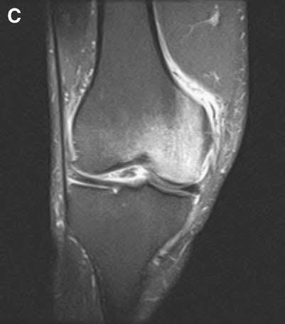
T2 / STIR MRI of the knee. Example image for illustration; marker positions are approximate.
Dr. Bora starts with your symptoms, how you move and your weight-bearing X-rays, and recommends an MRI only when it will guide your care.
Not every knee problem needs a replacement. Dr. Bora looks at whether joint preservation, arthroscopic repair or replacement is the safest and most effective choice for you and the way you live.
Keeps your natural jointTypical recovery
Protected weight-bearing for 2–3 weeks, then rehab over 6–12 weeks
Treat bone marrow lesions early to protect your natural knee.
Bone marrow lesions are areas of stress and swelling in the bone beneath the cartilage, and a common source of persistent knee pain. This treatment targets them directly, aiming to ease pain and slow joint breakdown so a replacement can be delayed or avoided.
Recovery times are estimates. Dr. Bora confirms the right option after examining you and reviewing your imaging.
A physician-led joint preservation pathway, not a one-size-fits-all procedure.
Examination and imaging guide every decision. Targeted subchondral treatment is considered only when it is appropriate, alongside strengthening, weight and load management, and follow-up.
Your weight-bearing X-rays, and an MRI if one is needed, are reviewed together to look at joint space, alignment and bone marrow lesions.
Learn why finding the right subchondral region matters, and how the full programme goes beyond a single procedure.

A subchondral injection is a minimally invasive treatment that places healing substances such as platelet-rich plasma (PRP) or adipocyte concentrate directly into the bone just beneath a joint’s cartilage.
Scroll to follow the procedure, step by step.
Step 1 of 4
TargetingYour MRI shows where the bone marrow lesion sits in the knee, in the spongy bone just beneath the cartilage. That area becomes the target.
Schematic illustration, not to scale. The exact technique, guidance method and substance used are decided by your surgeon for your knee. Response is variable, and pain relief is not assured.
Discuss my knee with Dr. BoraStandard knee surgery replaces the torn ligament—but ignores the violent bone impact underneath. In over 80% of ACL ruptures, the underlying subchondral bone sustains micro-fractures (bone marrow lesions) that cause persistent pain and trigger premature arthritis.
Arthrex Master Instructor
10,000+ Knee & ACL Procedures
“An ACL tear is not an isolated ligament injury. During the pivot-shift event, bone slams into bone, leaving a severe subchondral bone bruise that standard ACL surgery leaves completely untreated. By biologically treating the subchondral bone alongside ligament reconstruction, we protect the joint bed, accelerate graft osteointegration, and safeguard our athletes from post-traumatic arthritis.”
4 WAYS SUBCHOND™ TRANSFORMS ACL RECOVERY
During the pivot-shift event, the lateral femoral condyle violently impacts the posterior tibial plateau. This crushes microscopic trabeculae and pools fluid inside the subchondral bone marrow. Standard surgery only fixes the torn ligament—leaving this severe bone edema untouched to cause chronic pain.
Targeted subchondral adipocyte-cell delivery provides regenerative signaling cells and growth factors directly into the injured bone, accelerating trabecular healing and eliminating deep joint ache within weeks.
Have an MRI scan of your ACL or knee?
Dr. Manu Bora reviews MRI scans for subchondral bone bruises.
Why modern orthopaedics treats the bone bed alongside ligament reconstruction
| Clinical Factor | Standard ACL Surgery Only | SUBCHOND™ Augmented ACL Care |
|---|---|---|
| Torn ACL Ligament | Reconstructed with tendon graft | Reconstructed with anatomical precision |
| Subchondral Bone Bruise (BML) | Untreated (often lasts 12–24 months as chronic ache) | Targeted adipocyte injection heals bone trabeculae rapidly |
| Graft-to-Bone Tunnel Healing | Mechanical healing alone (risk of tunnel widening) | Biological stimulation accelerates osteointegration |
| 10-Year Osteoarthritis (PTOA) Risk | ~50% develop post-traumatic arthritis | Protects cartilage cushion from secondary degeneration |
| Return to High-Impact Sport | Delayed by lingering deep bone ache & stiffness | Accelerated confidence, zero residual bone edema |
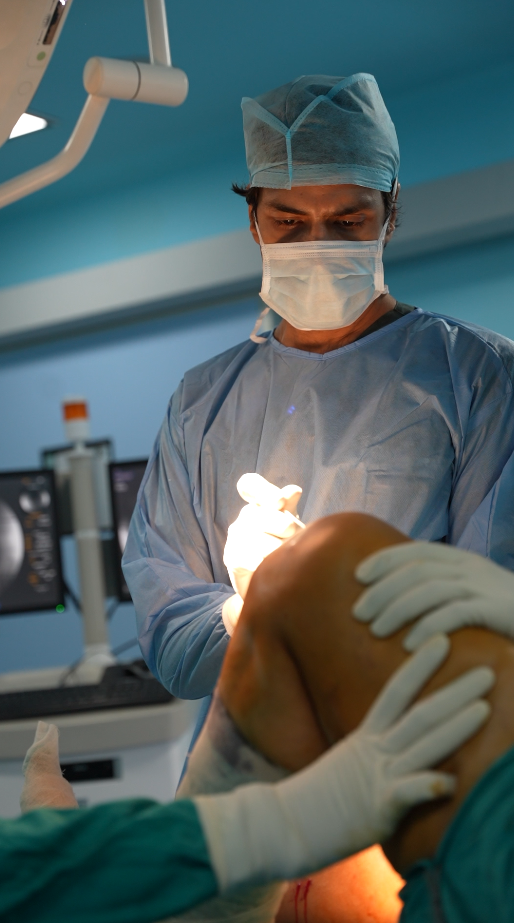
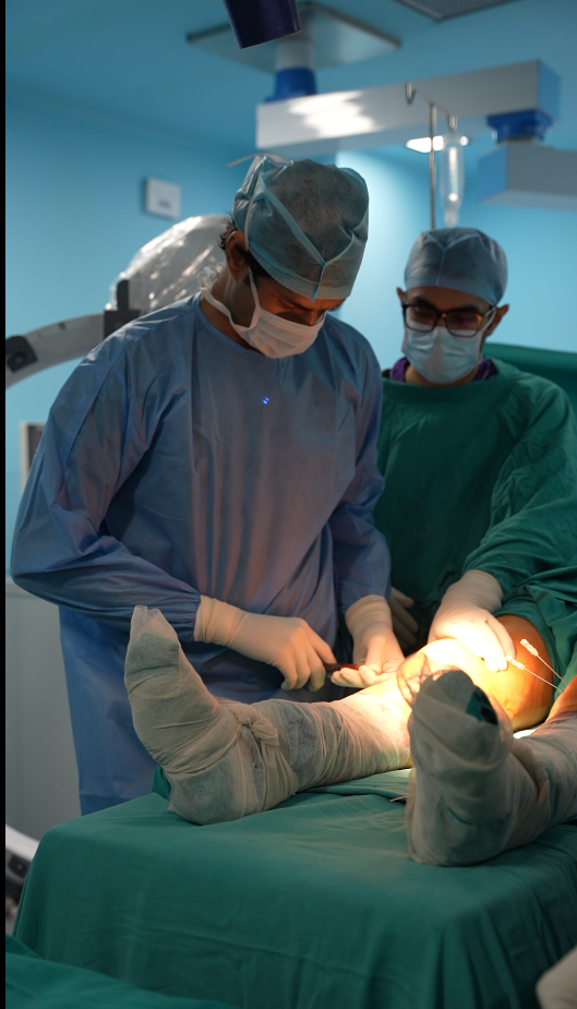
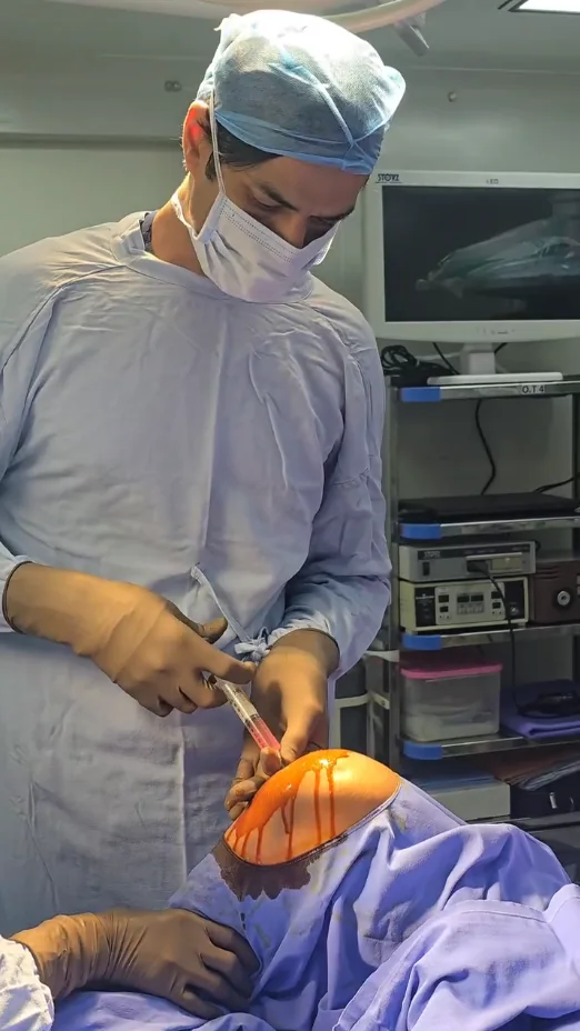
Photographs directly from our operating theatre. Demonstrating sterile preparation, imaging-assisted delivery, and surgical rigor.
Diagnosis, an appropriate clinical decision, guided rehabilitation, strength, recovery and thoughtful monitoring all belong together.
Diagnosis and clinical care
Dr. Manu Bora: examination, imaging review, and an appropriate discussion of joint preservation or replacement.
orthosport.in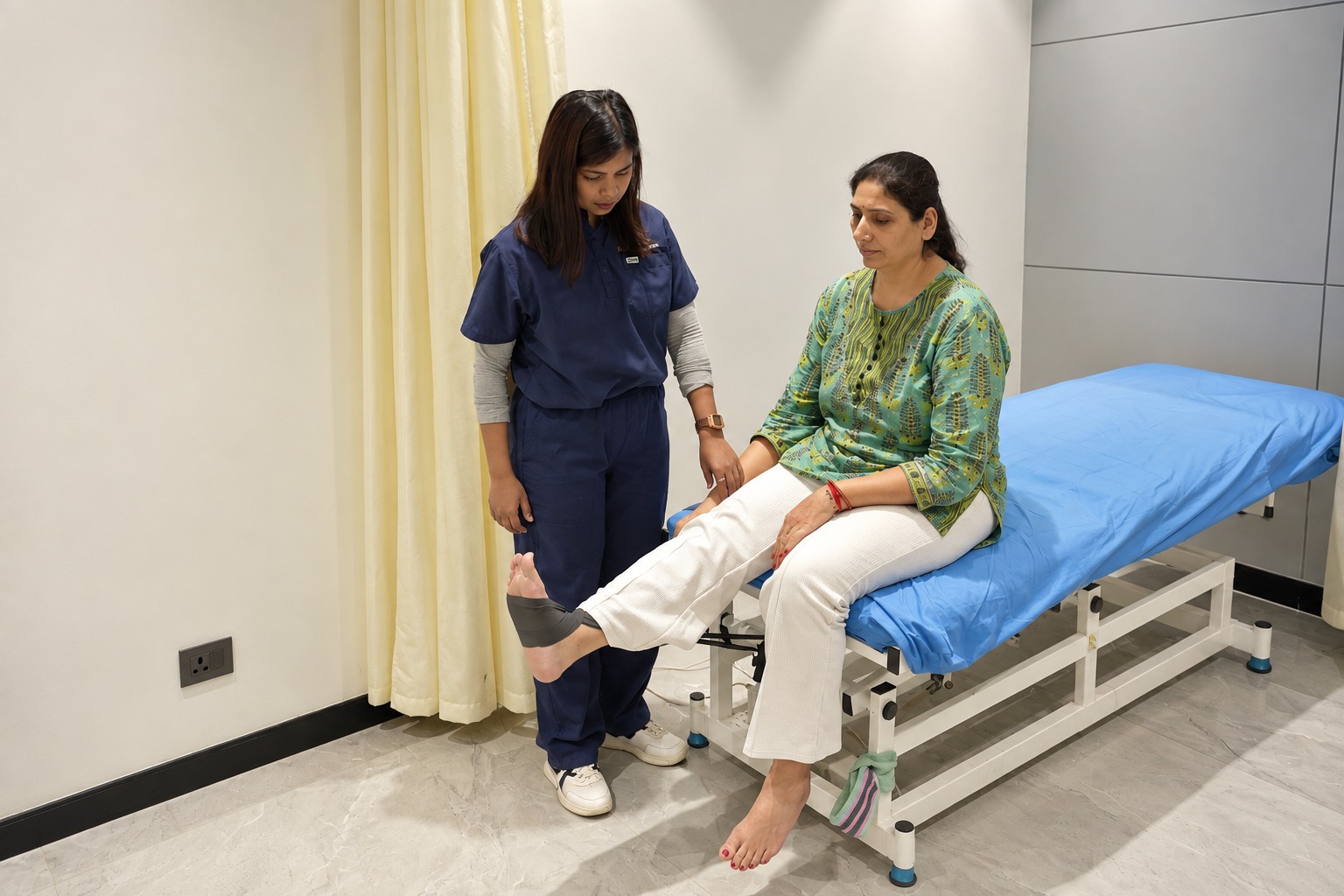
Rehab and strength
Individualised physiotherapy, regular check-ins and guided progression through in-person or online sessions.
threadsphysio.com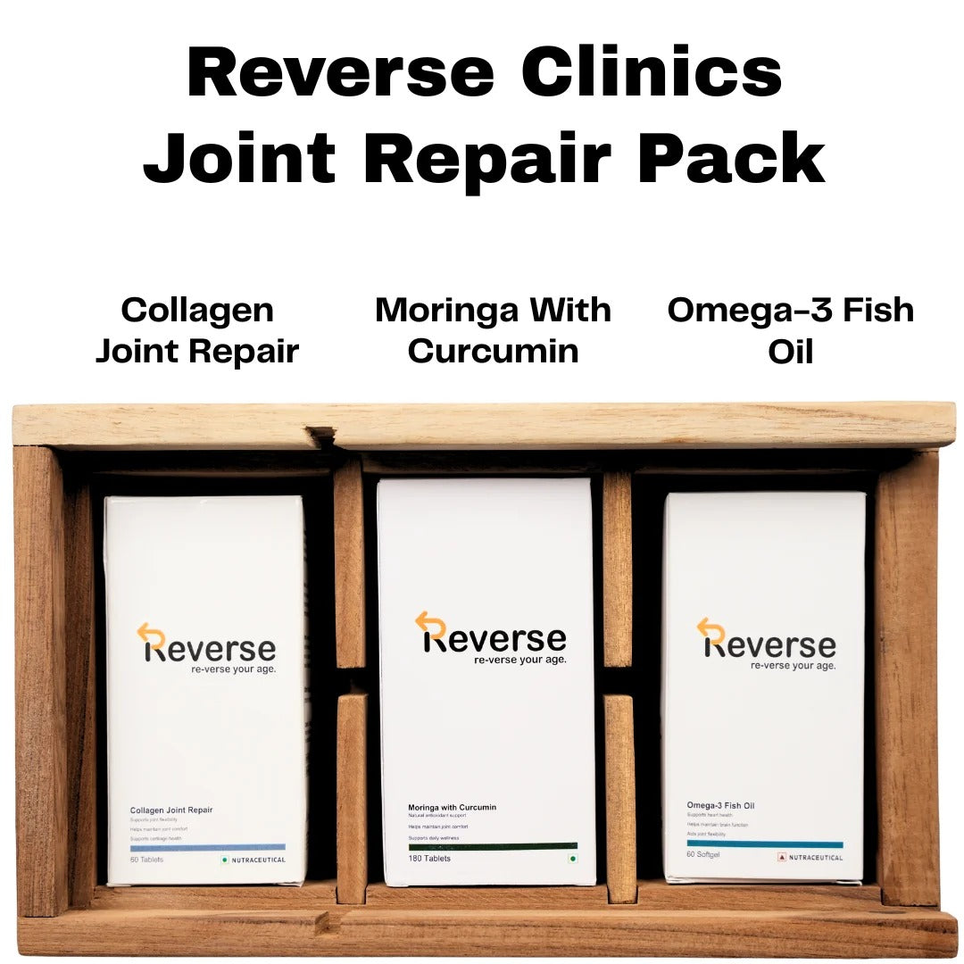
Recovery and lifestyle
Optional nutrition and supplement guidance where appropriate. No supplement replaces treatment or proves knee-regeneration claims.
reverseclinics.comResponse is variable, and pain relief after cell-based procedures is not assured. Progress also depends on disease severity, loading, rehabilitation and other factors.
Discuss my knee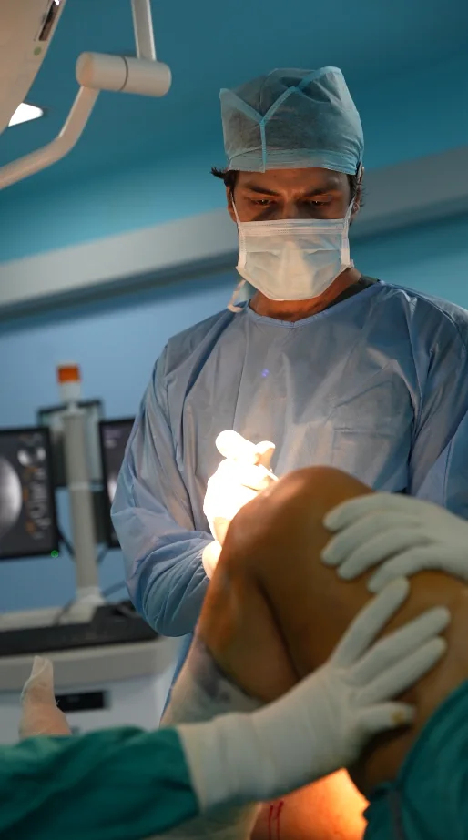
Early phase
Procedure-related soreness may need time to settle. Follow the mobility and load instructions provided.
Review window
Clinicians may review changes in pain, function and rehab progress. This is not a guaranteed benefit timeline.
Rehab and function
Progressive strength, walking tolerance and daily activity may continue to develop.
Long term
Monitor your symptoms, function, activity goals and imaging when clinically useful.
Clear, transparent answers to help you understand your knee condition, diagnostic options, and joint preservation pathways.
Our clinical team is available to evaluate your MRI scan or answer questions regarding your knee treatment options.
REQUEST AN EVALUATION →Book a personal consultation or request a second opinion on your MRI scan with Dr. Manu Bora at our centres in Gurugram, New Delhi, or Mumbai.
MS Orthopaedics · Arthrex Master Instructor · Sports Injury Specialist
Every consultation and MRI scan is reviewed directly by Dr. Manu Bora. You receive an honest, unbiased assessment whether your natural knee can be preserved.
Prior appointment only
Prior appointment only
Prior appointment only
Your privacy is respected. Clinical reports and medical data provided are strictly confidential and reviewed only by Dr. Manu Bora's surgical team.
Schedule a comprehensive orthopaedic evaluation with Dr. Manu Bora. Review your MRI scans, biomechanical alignment, and subchondral joint health.